Dental segmentation
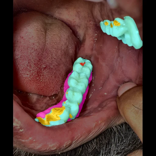
An analysis trained on 18,000+ intra-oral photos annotated by Dr Romain Lateur, collected in real clinical conditions.
Study carried out by iTransform GP · February 2026
18k+
Annotated photos
6
Shots per patient
8
Architectures evaluated
6
Clinical classes segmented
6 standardised shots per patient
Two arches, three views each — a reproducible protocol, annotated pixel by pixel by Dr Romain Lateur.
Maxillary

Left

Centre

Right
Mandibular

Left

Centre

Right
Le Voting Classifier
Rather than trusting a single model, MouthCare combines the predictions of five segmentation architectures through a pixel-level vote. Disagreement between models shows up as a lower probability, where a single model would decide without saying so.
- Pixel-level weighted vote across five segmentation architectures.
- UNet++, UNet, DeepLabV3+, PSPNet (ResNet-34 encoder) and SegFormer, retrained in October 2026.
- Segmentation into 6 clinical classes (teeth, gums, tartar, cavities…).
A demonstration on pre-recorded photographs. No photo can be sent from this page, and the result shown is neither a diagnosis nor medical advice.
MouthCare, in your pocket
Available on iOS and Android.